


- FAQs
- How To Get Help
- Microtia Treatment Options
Microtia Microtia is a birth deformity of the ear that occurs about one in 5,000-7,000 births, depending on varying statistics in different countries and in different ethnic races. When broken down in Latin, the term “microtia” is easy to … Continue reading
Microtia Microtia is a birth deformity of the ear that occurs about one in 5,000-7,000 births, depending on varying statistics in different countries and in different ethnic races. When broken down in Latin, the term “microtia” is easy to … Continue reading
Microtia Microtia is a birth deformity of the ear that occurs about one in 5,000-7,000 births, depending on varying statistics in different countries and in different ethnic races. When broken down in Latin, the term “microtia” is easy to … Continue reading
PATIENT RESULTS
About Microtia

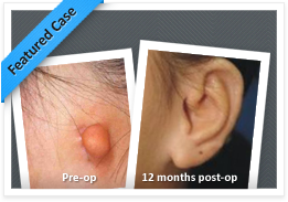
A small lobular type microtia.
Microtia
Microtia is a birth deformity of the ear that occurs about one in 5,000-7,000 births, depending on varying statistics in different countries and in different ethnic races. When broken down in Latin, the term “microtia” is easy to understand. Micro=small; Otia=ear.
Microtia is classified into four different types :
Grade I A slightly small ear with a shape much like a normal ear only smaller and usually a small but narrow ear canal.
Grade II A partial or hemi- ear with usually an absent ear canal
Grade III Absence of most of the external ear with a small “peanut” shaped remnant ear lobule and an absent ear canal and ear drum (atresia)
Grade IV Total absence of the ear or anotia
The most common grade is Grade III also known as lobular microtia.
Atresia
Atresia is absence or underdevelopment of the ear canal and middle ear structures. Microtia is almost always accompanied by atresia because the outer ear and the middle ear develop from one common block of tissue at the same time of development in the womb. Some microtia patients have what appears at first look to be a normal canal, but many of them are a “blind alley.” In contrast to atresia, canal stenosis is where there is an ear canal but it is much narrower than normal .Stenosis may be associated with a normal middle ear so the hearing will be normal or may have an underdeveloped middle ear causing severe conductive hearing loss. Even is the middle ear is normal in canal stenosis there are more problems with buildup of wax and difficulty cleaning so this in itself causes hearing loss and often warrants an attempt to widen the canal. The procedure to widen a narrow canal may oftne be much more simple than atresia surgery.
Causes of Microtia and Atresia
The exact cause (etiology) of microtia is unknown.
The incidence of microtia is one in 5-7,000. Parents who have a child with microtia then have an increased risk of 1 in 20 for subsequent children. The risk of a child with microtia eventually having a child of his/her own with microtia is also one in twenty.
The majority of patients with microtia have no other problems aside from the ear. Approximately 50% have underdeveloped bony and soft tissues on the involved side of the face (hemifacial microsomia) and 15 percent have facial nerve weakness. Other general abnormalities such as cleft lip, cardiac, or urological problems are encountered much less frequently.
Children with microtia become aware of their differences at about 3-4 years of age. Typically, they start comparing sides in a mirror and begin referring to their microtia as their “little ear “ but remain unconcerned by this difference. Awareness of facial difference between children starts around 5 years of age and an ear deformity may attract innocent curiosity at this age from peers. Teasing is almost unheard of in our experience with microtia but if it occurs it is not until 8-12 years of age. More commonly it is a desire to fit in that manifests around 10-12 years of age in most children with microtia and leads to a request for reconstruction. However close to 25% of patients live normal lives with their microtia/atresia and never request surgery.
Treatment Options
The two main methods for repairing microtia rely on different sources for the ear framework. One method uses a living sculpture from the patient’s own tissues (rib cartilage) and the other uses a framework of firm artificial plastic covered in a “living membrane” plus skin grafts (Medpor). This is the main difference between the two techniques: one type of ear is created from a patient’s own human tissues whereas the Medpor method combines foreign material and the patient’s own tissues.
Autogenous/Rib Graft Reconstruction
So far internationally this method in experienced hands remains the absolute gold standard for ear reconstruction as since the patient’s own tissues are used once an ear is “good” after a successful reconstruction it will remain good forever and never deteriorate over time. It also will be as resistant to the knocks and trauma of life as a normal ear and heal normally if injured. The rib graft method continues to be refined and improve over the years. In the 1970’s when it first started by Dr Radford Tanzer it involved 4-6 stages . Burt Brent improved it further to requiring around 4 stages and things have improved even further since. What was regarded as a great result in the 1970’s or 1980’s using the Tanzer/Brent method would no longer be accepted as a great result now. THis is partly due to the pioneering development of the method by both Francoise Firmin of Paris, France and Satoru Nagata of Tokyo, Japan. These doctors dramatically improved the method to a 2 stage technique that almost routinely produces good or excellent results in surgeons dedicated to the technique.
With the Nagata/Firmin method the first stage involves creating the ear framework and placing it under the skin where the ear should be on the scalp then at the second stage 3-6 months later the reconstruction is completed by elevating the ear from the side of the head using a flap of tissue and a skin graft.
Mr Andrew Greensmith continues to refine the Nagata method of reconstruction. In most cases with the use of exciting innovations in technique developed by Mr Greensmith ear reconstruction with rib cartilage now involves one main operation without the need for a later elevation procedure and so avoids any skin grafts. This new method involves either the placement of the small osmotic tissue expander 3-6 weeks before the main operation in order to grow more ear skin to cover a more projecting framework or creating the new skin by injecting hyaluronic acid gel ( the same product used to fill wrinkles and contour defects in cosmetic surgery ) serially over 8-12 weeks prior to the main operation. Both are novel forms of tissue expansion applied first to ear reconstruction for microtia by Mr Greensmith .
Ear reconstruction with rib cartilage can be undertaken once the patient’s chest circumference measures close to 60cm and this usually corresponds to an age of 8-10 onwards. In reality many 8-10 years olds are not ready psychologically for surgery as they are just not concerned enough to worry about their microtia deformity so tend to not start asking for a reconstruction until early teenage years. Some do not even bother asking until they are adults. Thankfully there is no upper limit to the age where a rib reconstruction can be performed. From 8-10 years of age a child can start to appreciate the difference in results that can be achieved by the rib graft method versus the Medpor method so can be involved in the decision making process.
In the method used by Mr Greensmith rib graft ear reconstruction involve a 1-2 night hospital stay after a 6-8 hour operation. Mr Greensmith uses the new method of rib restoration developed by Dr Nagata to rebuild the ribs harvested so that there is no noticeable donor sit defect or contour defect on the chest wall even in very thin patients. The Nagata method of rib graft harvest is also minimally painful as no muscle is cut to get to the ribs and this make a nice difference to the recovery process. In addition a Pain Buster catheter is place in the rib donor area beneath the muscles to deliver an infusion of local anaesthetic to the area for the first 24-48 hours after surgery. Mr Greensmith does not use drains in the chest or the ear reconstruction site. A small amount of scalp hair is shaved around the area for the ear reconstruction. A dressing protects the new ear for 2 weeks after which time it is removed and the patient can then leave it open and get their head wet in the shower.
See the “Results” section for a gallery of photos of rib graft reconstructions.
Medpor Ear Reconstruction
Medpor reconstruction uses a framework of artificial material called porous polyethythene made in one standard shape but a number of sizes. This implant was first used for ear reconstruction by Alexander Berghaus in Germany and he was the first to use the method combining the implant with a fascial flap and skin graft and continues to do so to this day. In the early 1990’s Dr John Reinisch a surgeon from the USA who had tried rib graft reconstruction but could not get it to work well also started to explore Medpor and began using it although initially he placed it directly under the ear/scalp skin and had terrible problems with implant exposure. When he later also started removing most of the ear deformity skin,using the additional fascial flap ( temporoparietal flap ) plus skin grafts he found his complication rate dropped and he was usually able to get it to work consistently. Despite Berghaus/Hempel of Germany and Reinisch in the USA having performed fairly large numbers of these reconstruction the majority of experts in ear reconstruction remain unconvinced by the method and feel even when it goes well that it gives an inferior result to rib graft with a poor colour match and the lifelong uncertainty of possible risks of implant fracture, trauma and exposure. However Mr Greensmith believes the technique does have merit in select cases or where the patient or parents insist on this method over the rib graft so long as the patient/parents understand that once a Medpor reconstruction is done you cannot convert it to a rib graft reconstruction.
Medpor reconstruction is marketed by it proponents as being able to be done as young as 2-3 years of age and creating “an ear before school”. Having extensive experience working with children of all ages at the Royal Children’s Hospital in Melbourne for over 10 years now Mr Greensmith believes that is is extremely unwise to submit a child at such a young age to such a complex procedure considering the child is given no part to play in making a choice between the rib method and the Medpor method let giving consideration to whether they even want their ear reconstruction. Assuming a child will cope badly with a deformity as minor as microtia is not exactly a pathway to building resilience in later life since any ear reconstruction will never exactly match the other normal ear in unilateral microtia and the child not the parents will live with the reconstruction good or bad , problematic or not , for the rest of their lives . Early surgery is much like operating on the parents and their anxieties , not the child.
Medpor surgery involve a 5-6 hour operation and a day stay or overnight procedure with no drains. A much larger portion of scalp is shaved compared to the rib graft method. Unlike the rib graft method a skin graft is used. Mr Greensmith take this entirely from the scalp to get the best colour match and avoid the 10-15 cm groin scar of the Reinisch method ( which is likely to grow pubic hair on the ear later in life ). Dressing are required for 2-4 weeks after surgery depending on the quality of graft take.
See the “Results” section of the this web site for a gallery showing representative Medpor results.
OUR LOCATION
REQUEST AN APPOINTEMENT
After submitting the form above, you will be contacted by our staff to answer any questions and discuss scheduling an appointment. You can also call us directly at +61 3 9508 9508 or Lee Dowling PA to Dr Greensmith on leed@melbplastsurg.com.